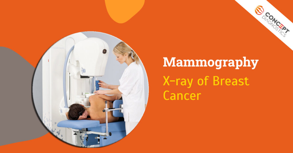
Mammography: X-ray of Breast
Mammograph is an X-ray image of breast. Mammography is delineated to capture images of breast to identify or detect any tissue abnormalities or lumps and tumor in breast of the patients. Moreover, it plays an important role to identify the signs and symptoms of breast cancer in patients.
Why is it done?
Mammography is usually done to serve two purposes;
Screening mammography: mostly, screening mammography is used to detect early cancer in women when they undergo no signs and symptoms of breast cancer. Timely screening mammogram helps in narrowing down the number of deaths occurred due to breast cancer.
Before experiencing any symptoms, it detects the growth of the cancerous tissues so can be taken measures for early treatments to avoid spreading. It exposes you to low-dose radiations but get you to know even if there any other abnormalities.
Diagnostic mammography: Usually, this is done when woman is undergoing any symptoms or signs such as lumps in breast, extreme pain in breast, discharge from nipple, unusual skin color, changed shaped or size of breast. This medical condition may not be cancer but it is essential to get tested to avoid risk.
Before Mammography:
- Pick out the best and certified diagnostic center for your test
- Discuss about your medical conditions (If any), pregnancy before test
- You can drink and eat as usual before test
- If you have undergone mammography prior, then bring old mammograms with you
- Avoid using deodorants, lotions, creams etc. before test
- Remove all the jewelry
- You will be given hospital gown to wear while performing this test
During Mammography:
- We will have you to stand in front of mammogram machine i.e., a device comprises of 2 surfaces, which compress the breast in between to detect abnormalities in patient’s breast.
- Then our technician will place your breasts on the lower surface and adjust the platform according to your height
- Your breasts will be compressed by the 2 plates by applying pressure to spread tissues of breast for few seconds.
- The applied pressure will keep the breast firm, hold and spread it evenly
- During radiation exposure, you need to hold your breath for few seconds and stand firmly
- It is little painful and may create discomfort to woman
- The breast image will be taken from 2 different angles in black and white color and captured on monitor.
After Mammogram
- Captured imaged will be checked by the technician, whether it is clear or not
- If it is still unclear, the process needs to be repeated
- You may feel mild discomfort or pain in your breast
- Within an hour, you can go back to your normal routine
- Your reports will be sent to the doctor for further probe
- Whole process takes about 30-40 minutes
Results:
You need to visit the clinic once you get the reports to discuss with the doctor;
- Cancerous growth
- Lumps in the breast
- Cysts
- Tumor
- Malignant tumor
- Calcium deposition in the ducts
If there are any unexplained abnormalities you may need biopsy or MRI to get a clear result. Sometimes mammography missed to capture some tissues or some issues. So, you may be advised to get for magnified test in order to get better understanding. At Concept Diagnostics, get tested and get detailed reports within short stretch of time.

